YouTube & Podcast
Everything Dr. Hill makes, in one place: the Head First podcast, weekly Neurofeedback & Chill livestreams, guest appearances, and channel videos. Use the filters to find what you want.
Subscribe on YouTube
📺Neuroscience insights, brain optimization tips, and live Q&A. New content weekly.
🔴Subscribe to @DrHillNeurofeedback & Chill
Every Monday • 6PM Pacific
Live neuroscience Q&A, brain training demos, and community discussion.
📺Watch Live Streams
Where Is Consciousness in the Brain? | Neurofeedback & Chill
In a major 2024 study, about one in four behaviorally unresponsive patients in a large convenience sample could still follow commands through patterns detected with EEG or fMRI. The finding exposes a basic problem: behavior, brain processing, and conscious experience can come apart. So where is consciousness in the brain? Tonight I separate wakefulness from conscious content, examine what anesthesia, covert consciousness, blindsight, neglect, and split-brain cases can actually tell us, and compare the major families of consciousness theory without pretending the field has selected a winner. A 2025 adversarial collaboration tested key predictions from global neuronal workspace theory and integrated information theory. The results challenged important parts of both. This is episode two of the Consciousness to Cadence series. Episode three asks whether an artificial system could satisfy any defensible evidence standard for consciousness. 🧠 KEY TAKEAWAYS: • Behavioral responsiveness is useful evidence about consciousness and an imperfect detector of it • In a 2024 six-center convenience sample, 60 of 241 unresponsive participants showed command-following on EEG or fMRI • Arousal and the contents of experience are related dimensions with partly separable mechanisms • Conscious processing appears to depend on distributed and recurrent interactions, not one known consciousness center • Blindsight and split-brain findings remain more contested than their textbook summaries suggest • Global workspace, integrated information, higher-order, recurrent processing, and predictive approaches make overlapping claims • A large 2025 study challenged key predictions of both GNWT and IIT and did not identify a winner • A neural correlate is not automatically the cause, full mechanism, or location of experience 📚 KEY RESEARCH: • Bodien et al. (2024). Cognitive Motor Dissociation in Disorders of Consciousness. New England Journal of Medicine. https://doi.org/10.1056/NEJMoa2400645 • Cogitate Consortium et al. (2025). Adversarial testing of global neuronal workspace and integrated information theories of consciousness. Nature. https://doi.org/10.1038/s41586-025-08888-1 • Seth and Bayne (2022). Theories of consciousness. Nature Reviews Neuroscience. https://doi.org/10.1038/s41583-022-00587-4 • Mashour et al. (2020). Conscious Processing and the Global Neuronal Workspace Hypothesis. Neuron. https://doi.org/10.1016/j.neuron.2020.01.026 • de Haan et al. (2020). Split-Brain: What We Know Now. Neuropsychology Review. https://doi.org/10.1007/s11065-020-09439-3 • Derrien et al. (2022). The nature of blindsight. Neuroscience of Consciousness. https://doi.org/10.1093/nc/niab043 RESOURCES: 📖 Neurofeedback: Explained: https://www.amazon.com/dp/B0GZ7QLM44 📞 Free consultation: https://www.peakbraininstitute.com/contact-us/locations 🌐 Dr. Hill's research: https://www.andrewhillphd.com/research #Consciousness #Neuroscience #EEG #Brain #AIConsciousness #DrAndrewHill #NeurofeedbackAndChill
What Happens in Your Brain When Neurofeedback Works? | Neurofeedback & Chill
What happens in the brain at the exact moment neurofeedback delivers a reward? My current preprint reanalyzes 64-channel EEG from a double-blind, active-placebo-controlled experiment to look at reward-locked brain activity, training frequency, and changes that remained at a follow-up visit about one month later. The most interesting result was a dissociation. Beta training produced the strongest observed reward-locked response, while both SMR groups showed the clearest durable growth in resting alpha. A matched baseline probe then split the reward-locked result into two parts. It confirmed that active reward timing selected elevated trained-band states relative to matched sham. It also showed that the current epoch cannot independently isolate an additional post-reward response from that selected state. Tonight I will walk through what we measured, what the sham controls, why a preprint still needs skepticism, and why physiology changing after an intervention does not automatically prove a clinical mechanism. I will also explain a real limitation in the reward-locked analysis that belongs in the interpretation. This is episode one of a six-part arc moving from brain measurement to consciousness, artificial intelligence, agency, ADHD, and the science of timing action. 🧠 KEY TAKEAWAYS: • EEG measures voltage differences at the scalp, not thoughts or a direct picture of consciousness • Neurofeedback is a closed loop: measure, translate, feed back, adapt, and measure again • The study included 40 healthy adults, three active protocols, active-placebo sham, five training sessions, and a follow-up visit • Active reward timing selected elevated trained-band states relative to band- and site-matched sham • The baseline probe preserved that manipulation check while narrowing the claim about a separate post-reward effect • Beta showed the strongest observed acute response while SMR showed the clearest durable resting-state change • Acute control and consolidation appear to be separable properties • The paper is a preprint, uses small groups, and does not contain clinical outcomes • Reward occurred after 500 milliseconds of threshold achievement, which complicates the interpretation of the post-reward decrease • A brain change can establish target engagement without establishing symptom efficacy 📚 KEY RESEARCH: • Hill (2026). Frequency-Specific Operant Learning in Neurofeedback Reveals Distinct Cortical Mechanisms. bioRxiv. https://doi.org/10.64898/2026.04.13.718260 • Sitaram et al. (2017). Closed-loop brain training: the science of neurofeedback. Nature Reviews Neuroscience. https://doi.org/10.1038/nrn.2016.164 • Ros et al. (2020). CRED-nf checklist. Brain. https://doi.org/10.1093/brain/awaa009 • Thibault and Raz (2017). The psychology of neurofeedback. American Psychologist. https://doi.org/10.1037/amp0000118 RESOURCES: 📖 Neurofeedback: Explained: https://www.amazon.com/dp/B0GZ7QLM44 📞 Free consultation: https://www.peakbraininstitute.com/contact-us/locations 🌐 Remote neurofeedback: https://www.peakbraininstitute.com/remote-neurofeedback-programs 🌐 Research and preprints: https://www.andrewhillphd.com/research #Neurofeedback #EEG #BrainTraining #Neuroscience #BrainPlasticity #DrAndrewHill #NeurofeedbackAndChill
The GLP-1 of Neurofeedback? A Brain Protocol for Cravings
In neurofeedback we often use a frontal EEG protocol that often seems to reduce grazing and compulsive snacking. Other clinicians describe close variants for appetite and repetitive behavior. Is this the GLP-1 of neurofeedback? Tonight I break down the Fp1-M2 protocol, what each frequency band may be doing, where it overlaps with GLP-1 appetite and reward circuitry, and what the human neurofeedback research actually shows. I also use screen addiction to separate three possible bottlenecks: cue-driven wanting, failed inhibition, and perseverative looping. Those patterns can look identical from the outside while pointing toward different neurofeedback strategies. A 2026 meta-analysis found a preliminary signal for reduced food craving and binge frequency. A 2024 sham-controlled study found an early signal for prefrontal neurofeedback and problematic internet use in children. A separate placebo-controlled study found that central SMR training improved laboratory inhibitory control in healthy adults. None of those studies tested this exact Fp1-M2 recipe, and no controlled trial validates the Fz-Pz alpha protocol for screen addiction. The goal is to separate three things cleanly: published evidence, clinical observation, and a plausible circuit model. 🧠 KEY TAKEAWAYS: • GLP-1 drugs and frontal neurofeedback are not the same intervention • They may affect different parts of an overlapping appetite, reward, and control network • The exact Fp1-M2 protocol is a clinical protocol, not a validated weight-loss treatment • Screen overuse can reflect cue-driven wanting, weak response inhibition, perseveration, or a mixture • A central SMR study supports an inhibitory-control rationale, but it was not a screen-addiction trial • Fz-Pz alpha is a clinic-derived strategy for rumination and perseveration, not a validated screen-addiction treatment • A 2026 meta-analysis found preliminary neurofeedback effects on craving and binge frequency across seven small randomized trials • The closest EEG studies partly match the high-beta inhibit and disagree on theta direction • Fp1-M2 is a voltage-difference channel, not a laser pointed at one brain structure • Better craving control is not the same thing as treating an eating disorder 📚 SECTIONS: • Why I call this the snacker protocol • What GLP-1 drugs do to appetite and reward circuits • Where frontal neurofeedback may meet the same network • The exact Fp1-M2 setup • One behavior, three possible bottlenecks • What the neurofeedback trials actually found • What other providers are doing • How to test a clinical observation without turning it into hype • Who should be cautious • Q&A KEY RESEARCH CITED: • Chen et al. (2026). Seven-trial meta-analysis of neurofeedback for food craving and binge eating. *Psychiatry Research*. https://doi.org/10.1016/j.psychres.2026.116979 • Renaud et al. (2026). Sham-controlled dlPFC neurofeedback study protocol for food addiction and severe obesity. *Clinical Nutrition ESPEN*. https://doi.org/10.1016/j.clnesp.2026.103589 • Hilbert et al. (2024). Exploratory EEG and fNIRS neurofeedback trial for binge-eating disorder. *Psychological Medicine*. https://doi.org/10.1017/S0033291723002350 • Blume et al. (2022). Randomized pilot of EEG neurofeedback for binge-eating disorder. *Neurotherapeutics*. https://doi.org/10.1007/s13311-021-01149-9 • Kohl et al. (2019). Real-time fMRI neurofeedback of left dlPFC activity and eating behavior. *NeuroImage*. https://doi.org/10.1016/j.neuroimage.2019.02.033 • van Bloemendaal et al. (2014). Human GLP-1 receptor activation and food-cue responses. *Diabetes*. https://doi.org/10.2337/db14-0849 • Alhadeff et al. (2012). Endogenous GLP-1 projections into mesolimbic reward circuitry. *Endocrinology*. https://doi.org/10.1210/en.2011-1443 • Choi et al. (2024). Mobile prefrontal neurofeedback for problematic internet use in children. *Neuropsychiatric Disease and Treatment*. https://doi.org/10.2147/NDT.S454881 • Dousset et al. (2024). C3 SMR neurofeedback and inhibitory control. *International Journal of Clinical and Health Psychology*. https://doi.org/10.1016/j.ijchp.2024.100501 • Gu et al. (2025). VTA real-time fMRI neurofeedback in young adults at elevated gaming-disorder risk. *Journal of Medical Internet Research*. https://doi.org/10.2196/64687 • Klausen et al. (2026). Semaglutide for alcohol use disorder with obesity. *The Lancet*. https://doi.org/10.1016/S0140-6736(26)00305-3 RESOURCES: 🧠 Peak Brain Institute: https://peakbraininstitute.com 📖 *Neurofeedback: Explained*: https://www.amazon.com/dp/B0GZ7QLM44 🌐 Peak Brain Press: https://www.peakbrainpress.com 📚 Dr. Hill's articles: https://www.andrewhillphd.com/articles 🎥 GLP-1s Are Dopamine Drugs (Sort Of): https://www.youtube.com/watch?v=Q7CRCibfPrc 🎥 GLP-1s, ADHD, & OCD: https://www.youtube.com/watch?v=mRRJ3afCAv4 #Neurofeedback #GLP1 #FoodNoise #Cravings #BrainTraining #ScreenAddiction #Impulsivity #BrainHealth #QEEG
Does Addiction Age the Brain? Why Relief Becomes Need
A new July 2026 brain imaging study found older appearing structural MRI patterns in groups with alcohol use disorder, tobacco use disorder, or both. That is a real result, though not proof that addiction adds a fixed number of years to a person's brain. In this video, we will explore what the researchers actually measured, why pleasure can shift into relief, and how the brain can learn to need a state that a substance or behavior once supplied easily. The episode also marks the release of my new book *Borrowed Peace: The Recovery Guide for Brains That Self-Medicate*. Available now: https://www.amazon.com/Borrowed-Peace-Recovery-Self-Medicate-Different/dp/1971402028/ 🧠 KEY TAKEAWAYS: • Brain age is a model based on structural MRI patterns rather than a literal biological clock • The July study found group differences in alcohol and tobacco use disorders, but it cannot establish causation • The addiction samples were much smaller than the healthy training sample • As dependence develops, reward can weaken while stress and withdrawal systems become more reactive • Wanting and liking can separate • Self medication is one of several pathways into problematic use • Moderation and abstinence are clinical decisions shaped by substance, severity, safety, history, preference, and outcome • Sudden alcohol withdrawal after prolonged heavy drinking can be dangerous and may require medical care 📚 SECTIONS,: • The new brain aging result • What a brain age model can and cannot say • Why pleasure can become relief • Wanting after liking fades • What self medication gets right and wrong • The case for an honest menu of recovery goals • A practical regulation audit • What recovery can build • Live Q and A KEY RESEARCH CITED: • Liang et al. (2026). Brain aging patterns among nine neurological disorders: A case-control study. PLOS Medicine. DOI: 10.1371/journal.pmed.1004860 • Vendruscolo and Koob (2026). Neurobiology of negative reinforcement as a driving force in alcohol addiction. Neuron. DOI: 10.1016/j.neuron.2026.04.038 • Robinson and Berridge (2025). The Incentive-Sensitization Theory of Addiction 30 Years On. Annual Review of Psychology. DOI: 10.1146/annurev-psych-011624-024031 • Peart and Murray (2026). What's next for interoception and addiction? Neuroscience and Biobehavioral Reviews. DOI: 10.1016/j.neubiorev.2026.106875 • Bowen et al. (2014). Mindfulness-based relapse prevention, relapse prevention, and treatment as usual for substance use disorders. JAMA Psychiatry. DOI: 10.1001/jamapsychiatry.2013.4546 RESOURCES: 🧠 Peak Brain Institute: https://peakbraininstitute.com 🌐 Remote Neurofeedback: https://peakbraininstitute.com/remote-neurofeedback-programs 📖 Borrowed Peace: https://www.amazon.com/Borrowed-Peace-Recovery-Self-Medicate-Different/dp/1971402028/ 🍷 NIAAA Alcohol Treatment Navigator: https://alcoholtreatment.niaaa.nih.gov 🆘 FindTreatment.gov: https://findtreatment.gov #Addiction #BrainHealth #AlcoholUseDisorder #NicotineAddiction #Recovery #Neuroscience #WellnessMonth #BorrowedPeace #DrAndrewHill #NeurofeedbackAndChill
The Alzheimer's Sleep Paradox: When More Sleep Isn't Better
A new Alzheimer's study reports that early amyloid pathology can keep the brain from settling into normal NREM sleep, but the experiment was in mice. At the same time, new human studies link unusually long sleep with tau biomarkers, Alzheimer's-like brain atrophy, and poorer cognition. In this video, we separate cause from warning signal, explain the role of microglia and glymphatic clearance, and show why the right question is not simply, "How many hours did you sleep?" 🧠 KEY TAKEAWAYS: • The new microglia result comes from a mouse mechanism study • Early amyloid pathology reduced NREM sleep through reactive microglia even before plaque burden became severe • Long self-reported sleep tracks with Alzheimer's-related biomarkers in some human cohorts, but it may be a marker of early disease or fragmented sleep • Normal human NREM physiology appears to support overnight movement of amyloid beta and tau from brain to plasma • Sleep duration, sleep architecture, sleep quality, and time in bed are different measurements • Do not deliberately restrict needed sleep; investigate a persistent change in sleep need or daytime function KEY RESEARCH CITED: • Constantino et al. (2026). Reactive microglia drove plaque-associated NREM sleep loss in APP/PS1 mice. Alzheimer's & Dementia. DOI: 10.1002/alz.71579 • Young et al. (2026). Long self-reported sleep showed a nonlinear association with plasma p-tau181 in 2,410 Framingham participants. Alzheimer's & Dementia. DOI: 10.1002/alz.71499 • Anagnostakis et al. (2026). Sleep duration showed U-shaped associations with an Alzheimer's-like MRI signature and cognition in 38,816 UK Biobank participants. Alzheimer's & Dementia. DOI: 10.1002/alz.71528 • Dagum et al. (2026). A randomized crossover study supported sleep-active movement of amyloid beta and tau from brain to plasma in humans. Nature Communications. DOI: 10.1038/s41467-026-68374-8 #Alzheimers #Sleep #BrainHealth #Dementia #GlymphaticSystem #Microglia #Neuroscience #HealthyAging #DrAndrewHill #NeurofeedbackAndChill **Resources / Work with Dr. Hill:** 📞 Free consultation: https://calendly.com/drhill1 🧠 Peak Brain Institute: https://peakbraininstitute.com 📖 Dr. Hill's articles and books: https://www.andrewhillphd.com/
Brain Criticality & ADHD: Flexibility, Hyperfocus, and Losing the Thread
ADHD is labeled as if your attention is missing. But people with ADHD can hyperfocus for hours, respond brilliantly under pressure, and then fail to start a simple email. The problem is not the existence of attention. It is controlling the state attention is in, and moving that state when the task changes. A study published today in *Translational Psychiatry* offers a new systems-level explanation. Researchers recorded MEG activity from adults with and without ADHD during rest and two continuous-performance tasks. They measured long-range temporal correlations: whether changes in brain oscillations carry structured memory across seconds and minutes. The ADHD group showed stronger and differently task-modulated correlations, while symptom severity tracked where individuals appeared to sit within a "critical" regime between activity that dies out too easily and activity that propagates too strongly. In this livestream, I connect that result to 25 years of QEEG work, the three ADHD biotypes we previously talked about that were identified in a 2026 *JAMA Psychiatry* study, the GO/STOP attention model in *Gifted & Tortured*, ADHD sleep and arousal problems, hyperfocus, stimulants, and neurofeedback. The critical question is not "does an ADHD brain have too much or too little activity?" It is "can this brain enter the right state, hold it, and leave it when the environment changes?" 🧠 **KEY TAKEAWAYS:** • Brain criticality is a complex-systems concept—not a medical emergency and not simply "more brain activity" • The study recruited 34 adults with ADHD and 36 controls and used MEG during rest, visual detection, and Go/NoGo tasks • ADHD participants did not perform worse on most measures; the clearest behavioral difference was more false alarms, suggesting impaired braking or compensation rather than global cognitive failure • Brain dynamics differed even when average behavior looked similar: ADHD participants showed stronger long-range temporal correlations and different rest-to-task modulation • Symptom relationships were not uniformly linear. Moderate traits sometimes sat near the putative functional sweet spot, while greater severity was associated with movement toward a more excitation-dominated state • This may help reconcile ADHD impairment with hyperfocus, novelty seeking, cognitive flexibility, and context-dependent strengths • The paper does not create an ADHD diagnostic test, prove a dopamine theory, or demonstrate that neurofeedback changes criticality • The practical target remains state regulation: sleep, task structure, movement, medication when appropriate, and individualized—not diagnosis-only—brain training **Research and related reading:** • Haque et al. (2026), *Translational Psychiatry*: https://doi.org/10.1038/s41398-026-04267-0 • Pan et al. (2026), three ADHD biotypes in *JAMA Psychiatry*: https://doi.org/10.1001/jamapsychiatry.2026.0001 • Westwood et al. (2025), ADHD neurofeedback meta-analysis: https://doi.org/10.1001/jamapsychiatry.2024.3702 • ADHD Brain Types: https://www.andrewhillphd.com/articles/adhd-brain-types • Does Neurofeedback Work for ADHD?: https://www.andrewhillphd.com/articles/neurofeedback-adhd-guide • Why Does My ADHD Kid Make Me Yell?: https://www.andrewhillphd.com/articles/adhd-parenting-yelling **Research and related reading:** • Haque et al. (2026), *Translational Psychiatry*: https://doi.org/10.1038/s41398-026-04267-0 • Pan et al. (2026), three ADHD biotypes in *JAMA Psychiatry*: https://doi.org/10.1001/jamapsychiatry.2026.0001 • Westwood et al. (2025), ADHD neurofeedback meta-analysis: https://doi.org/10.1001/jamapsychiatry.2024.3702 • ADHD Brain Types: https://www.andrewhillphd.com/articles/adhd-brain-types • Does Neurofeedback Work for ADHD?: https://www.andrewhillphd.com/articles/neurofeedback-adhd-guide • Why Does My ADHD Kid Make Me Yell?: https://www.andrewhillphd.com/articles/adhd-parenting-yelling **Work with Dr. Hill:** 📞 Free consultation: https://calendly.com/drhill1 🧠 Peak Brain Institute: https://peakbraininstitute.com 📖 Dr. Hill's articles and books: https://www.andrewhillphd.com/ #ADHD #BrainCriticality #Neuroscience #BrainHealth #Attention #ExecutiveFunction #Neurofeedback #QEEG #DrAndrewHill #NeurofeedbackAndChill
Are Microplastics Really in Your Brain? Maybe. Maybe not..
Scientists have reported microplastics in human blood, lungs, arterial plaques, and even the brain. Then a 2026 laboratory study found that ordinary nitrile and latex gloves can deposit thousands of particles that standard instruments misidentify as polyethylene. A second measurement problem is even stranger: fatty human tissue can produce the same chemical fragments as plastic when it is heated for analysis. So is the entire microplastics story wrong? Probably not completely. But the field has been collapsing four separate claims into one: exposure, detection, accumulation, and harm. Tonight I separate those claims, show which human findings survive the new criticism, and examine the emerging market for plasma exchange, supplements, and other supposed microplastic detoxes. The real story is neither "plastic is poisoning every brain" nor "it was all the gloves." It is a young field trying to measure an almost impossibly contaminable target. 🧠 KEY TAKEAWAYS: • The 2026 glove study is real, but it does not erase every microplastic finding. It most directly undermines small particles identified as polyethylene by spectral matching • A separate problem affects pyrolysis testing: fats and waxes can generate polyethylene-like chemical fragments, especially in lipid-rich tissue such as the brain • Human exposure is certain and tissue deposition is probable. The most credible signals come from larger, non-polyethylene particles found above procedural blanks in lungs and olfactory tissue • Detection is not the same as accumulation, and accumulation is not the same as demonstrated harm • No treatment has been shown to remove microplastics from human tissues or improve health by doing so • Plasma exchange is an interesting experiment, not a validated detox. In the newest study, the plastic tubing apparently added particles to people who started with low readings • Reducing exposure is more evidence-grounded than trying to extract particles after they enter the body 📚 SECTIONS: • Four claims hiding inside one frightening headline • The glove error that fooled three major testing methods • The human evidence that survives the criticism • What the brain and dementia findings can and cannot show • Can plasma exchange, chitosan, blood donation, or sauna remove plastic? • What is reasonable to do while the science catches up? • Live Q&A KEY RESEARCH CITED: • Clough et al. (2026). Dry nitrile and latex glove contact produced a mean of approximately 2,000 microplastic false positives per square millimeter. Analytical Methods. DOI: 10.1039/D5AY01801C • Witzig et al. (2020). Stearates and fatty acids from ten disposable gloves were misidentified as polyethylene using Raman, FTIR, and pyrolysis-GC/MS. Environmental Science & Technology. DOI: 10.1021/acs.est.0c03742 • Tan et al. (2026). Biological fatty acids and wax produced polyethylene-like pyrolysis products and false-positive PE identifications. Microplastics and Nanoplastics. DOI: 10.1186/s43591-026-00185-6 • Jenner et al. (2022). Microplastics were detected above procedural blanks in 11 of 13 human lung samples. Science of the Total Environment. DOI: 10.1016/j.scitotenv.2022.154907 • Amato-Lourenço et al. (2024). Sixteen particles or fibers, mostly polypropylene, were detected in the olfactory bulbs of 8 of 15 decedents. JAMA Network Open. DOI: 10.1001/jamanetworkopen.2024.40018 • Nihart et al. (2025). Reported increasing microplastic concentrations in decedent human brains, with higher readings in dementia samples. Nature Medicine. DOI: 10.1038/s41591-024-03453-1 • Weinstein et al. (2026). Plasma exchange reduced proprietary blood-test readings at high starting levels but increased them at low starting levels, apparently because of plastic tubing. Journal of Clinical Apheresis. DOI: 10.1002/jca.70135 RESOURCES: 📞 Book a free consultation: https://calendly.com/drhill1 🧠 Peak Brain Institute: https://peakbraininstitute.com 🌐 Remote Neurofeedback: https://peakbraininstitute.com/remote-neurofeedback-programs 📖 Dr. Hill's articles: https://www.andrewhillphd.com/articles #Microplastics #Nanoplastics #BrainHealth #Neuroscience #Dementia #Detox #Neurofeedback #DrAndrewHill #NeurofeedbackAndChill #HealthResearch #ScienceNews
Your Brain on Hormones: Alzheimer's, Your Cycle, PMDD, & Menopause
If your brain feels like it changes through the month, sharper some weeks, foggier in the days before your period, you're not imagining it. And if you've hit your 40s and the fog stopped lifting, that's not in your head either. Estrogen and progesterone aren't just reproductive hormones. They're master neuromodulators of the cortex, and when they move, the brain moves with them, measurably, on the EEG. In this video we trace hormones and the brain across the whole female lifespan: how your cycle shifts your alpha rhythm, how PMDD pulls frontal alpha asymmetry into a depression-type pattern, and what happens to the brain at menopause. The through-line: these shifts are individual, which is exactly why you measure instead of assume. 🧠 KEY TAKEAWAYS: • Estrogen activates and builds (serotonin, dopamine, acetylcholine, plasticity, glucose metabolism, blood flow, BDNF); progesterone, via its metabolite allopregnanolone, a GABA-A neurosteroid, calms and inhibits. Two hormones, opposite jobs, both tuning the cortex. • Your resting EEG shifts across the menstrual cycle: peak alpha frequency runs highest in the luteal phase and lowest in the late follicular phase; alpha power tends to drop and theta to rise approaching ovulation. • These cycle effects are real but heterogeneous. They show up clearly in some women and barely in others. That individual variability is the whole point: you measure, you don't assume. • PMDD isn't "bad PMS." Women with PMDD show a depression-type frontal alpha asymmetry that emerges specifically in the luteal phase, and frontal alpha asymmetry is a trainable neurofeedback target. • The leading model of PMDD is abnormal CNS sensitivity to a normal hormonal shift (allopregnanolone/GABA-A), not abnormal hormone levels. • Menopause withdraws estrogen's multi-system support at once. Mosconi's imaging shows the transition is a critical window for long-term brain health. HRT timing, exercise, sleep, diet, and neurofeedback each help. 📚 Topics • Estrogen and progesterone: the brain's master neuromodulators • The monthly tide, your cycle on the EEG (alpha, IAF, theta) • When the tide pulls too hard, PMDD and frontal alpha asymmetry • The big shift, perimenopause, menopause, and what helps • Closing + Peak Brain CTA (Brain Awareness Month finale) Research Cited: • Brötzner et al. (2014). Peak alpha frequency highest in luteal, lowest in late follicular phase. Brain Research. DOI: 10.1016/j.brainres.2014.06.034 • García-Granados et al. (2026). Systematic review: alpha decreases, theta increases in late follicular phase, but literature is heterogeneous. Arch Womens Ment Health. DOI: 10.1007/s00737-025-01654-2 • Accortt & Allen (2006). PMDD shows depression-type frontal alpha asymmetry emerging in the luteal phase. J Abnorm Psychol. DOI: 10.1037/0021-843X.115.1.179 • Schmidt et al. (2017). PMDD symptoms are triggered by the normal cyclical change in ovarian steroids, not by abnormal hormone levels (CNS-sensitivity model). Am J Psychiatry. DOI: 10.1176/appi.ajp.2017.16101113 • Bäckström et al. (2021). Randomized trial of the neurosteroid sepranolone for PMDD; targets the allopregnanolone/GABA-A mechanism. Psychoneuroendocrinology. DOI: 10.1016/j.psyneuen.2021.105426 • Mosconi et al. (2017a). Sex differences in Alzheimer risk. Neurology. DOI: 10.1212/WNL.0000000000004425 • Mosconi et al. (2017b). Perimenopause and emergence of an Alzheimer's bioenergetic phenotype. PLoS One. DOI: 10.1371/journal.pone.0185926 • Maki & Henderson (2012). HRT critical-window hypothesis for cognition. Climacteric. DOI: 10.3109/13697137.2012.660613 RESOURCES: 📞 Book a free consultation: https://calendly.com/drhill1 🧠 Peak Brain Institute: https://peakbraininstitute.com 🧠 QEEG brain mapping: https://peakbraininstitute.com/brain-mapping 🌐 Remote Neurofeedback: https://peakbraininstitute.com/remote-neurofeedback-programs 📖 Dr. Hill's books: https://www.peakbrainpress.com and QUIZ: https://www.giftedandtortured.com/ #Hormones #MenstrualCycle #PMDD #Menopause #Perimenopause #BrainFog #Estrogen #QEEG #Neurofeedback #DrAndrewHill #NeurofeedbackAndChill #Alzheimers
The Vaccine Dementia Link: What the Data Shows
Can an ordinary shingles shot lower your risk of dementia? In 2025, one of the cleanest studies in years said maybe. People who got the live shingles vaccine were about 20% less likely to be diagnosed with dementia over the next seven years. The reason to care isn't just the result. It's how the study was built. Wales rolled out the vaccine with a strict birthday cutoff, and that accident of policy created a natural experiment almost as clean as a randomized trial. In this video, I break down the Welsh natural experiment, the infection-and-inflammation theory of Alzheimer's, and, most importantly, how to tell a strong causal claim from a weak one, so you can read health news without getting fooled. 🧠 KEY TAKEAWAYS: • Wales rolled out the shingles vaccine in 2013 with a strict birthdate cutoff, and that accident of policy created a natural experiment nearly as clean as a randomized trial • Eyting, Geldsetzer et al. (2025, Nature): the live shingles vaccine cut new dementia diagnoses by about 3.5 percentage points over 7 years, roughly a 20% relative reduction, stronger in women • The design (regression discontinuity) beats ordinary "vaccinated people get less dementia" studies because it removes healthy-vaccinee bias. The birthday cutoff acts like a coin flip • The leading mechanism, viral reactivation and neuroinflammation (VZV, HSV-1) feeding Alzheimer's pathology, is plausible but NOT proven; the vaccine may also work through non-specific "trained immunity" • This is not advice to get a shingles shot to prevent dementia. Talk to your doctor. But it is a master class in telling a strong causal claim from a weak one 📚 IN THIS EPISODE: • Can a vaccine actually lower dementia risk? • The Welsh natural experiment and what it found • How to tell a strong causal claim from a weak one • The mechanism: infection, inflammation, and amyloid • What this means for you, and the one question to ask any health headline KEY RESEARCH CITED: • Eyting, Geldsetzer et al. (2025). Shingles vaccine cut dementia diagnoses ~20% in a Welsh natural experiment. Nature, 641, 438–446. DOI: 10.1038/s41586-025-08800-x • Taquet et al. (2024). Recombinant (Shingrix) vaccine linked to lower dementia risk than the older live vaccine. Nature Medicine, 30, 2777–2781. DOI: 10.1038/s41591-024-03201-5 • Xie, Eyting, Geldsetzer et al. (2025). Shingles vaccination linked to slower progression across dementia stages and lower dementia mortality. Cell, 188, 7049–7064. DOI: 10.1016/j.cell.2025.11.007 • Moir, Lathe & Tanzi (2018). Amyloid-β as antimicrobial peptide / antimicrobial protection hypothesis. Alzheimer's & Dementia, 14, 1602–1614. DOI: 10.1016/j.jalz.2018.06.3040 • Kumar et al. (2016). Amyloid-β entraps microbes (preclinical support for the antimicrobial hypothesis). Science Translational Medicine, 8, 340ra72. DOI: 10.1126/scitranslmed.aaf1059 • Itzhaki (2018). Herpes simplex virus (HSV-1) hypothesis of Alzheimer's. Frontiers in Aging Neuroscience, 10, 324. DOI: 10.3389/fnagi.2018.00324 RESOURCES: 📞 Book a free consultation: https://calendly.com/drhill1 🧠 Peak Brain Institute: https://peakbraininstitute.com 🌐 Remote Neurofeedback: https://peakbraininstitute.com/remote-neurofeedback-programs 📖 Dr. Hill's articles: https://www.andrewhillphd.com/articles 📚 BOOKS (Peak Brain Press): https://peakbrainpress.com • Gifted & Tortured: your brain's phenotypes, and how they make you gifted and tortured. Out now. • Half a Second More: The Meditation Guide for Brains That Won't Sit Still. Signed pre-order copies available now from the author; releases June 30. #Dementia #Alzheimers #ShinglesVaccine #BrainHealth #Neuroinflammation #DrAndrewHill #NeurofeedbackAndChill #Neuroscience #CausalInference #BrainAwarenessMonth #EndALZ #Neurofeedback
There's Finally a Blood Test for Alzheimer's. Should You Get It?
For decades, getting a real answer about Alzheimer's meant a spinal tap, a PET scan, or waiting until the symptoms were obvious. As of 2025 there's a fourth option: a single blood draw. The FDA cleared the first one in May, and by August you could order it from a national lab. In this video we break down what the test actually measures, who it's really for (and who it's not), the caveats nobody mentions, and the one question it can't touch - how your brain is actually functioning right now. 🧠 KEY TAKEAWAYS: • The FDA cleared the first blood test for Alzheimer's: the Lumipulse G pTau217/β-Amyloid 1-42 Plasma Ratio (Fujirebio) on May 16, 2025. It's a diagnostic aid, not a screening test. • It's intended for adults 55+ who already have signs or symptoms of cognitive decline, evaluated in a clinical setting, NOT for healthy, asymptomatic people who feel fine and want to peek around the corner. • p-tau217 is the standout blood biomarker. The test's ratio agrees with amyloid PET about 92% of the time for positives and about 97% for negatives. • A positive result means amyloid is present - it does NOT by itself mean you have Alzheimer's. Amyloid shows up in cognitively normal people too, and kidney function can shift the number. • The blood test measures pathology: is excess amyloid there? A QEEG brain map measures function - how is your brain actually working? Those are different questions, and function is the trainable one. KEY RESEARCH CITED: • U.S. FDA (May 16, 2025). First blood test cleared to aid Alzheimer's diagnosis: Lumipulse G pTau217/β-Amyloid 1-42 Plasma Ratio (Fujirebio Diagnostics). In vitro diagnostic, 510(k), Breakthrough Device. Validation: multicenter study of 499 plasma samples from cognitively impaired adults vs amyloid PET/CSF with 91.7% positive percent agreement, 97.3% negative percent agreement, under 20% indeterminate. • Palmqvist et al., Nature Medicine (2025)" SEPARATE multicenter study of 1,767 participants with cognitive symptoms on a fully automated plasma p-tau217 platform; ~85% accuracy in primary care. DOI 10.1038/s41591-025-03622-w (PMID 40205199). • Ashton et al., JAMA Neurology (2024): plasma p-tau217 immunoassay establishes p-tau217 as the standout blood biomarker for Alzheimer's pathology. DOI 10.1001/jamaneurol.2023.5319. • Labcorp (Aug 18, 2025): nationwide launch of the FDA-cleared plasma p-tau217/β-amyloid 1-42 ratio test. • U.S. FDA (Aug 2025): subcutaneous lecanemab autoinjector (Leqembi IQLIK) approved for at-home maintenance dosing; lecanemab (Leqembi) and donanemab (Kisunla) approved for MCI/mild AD. RESOURCES: 📞 Book a free consultation: https://calendly.com/drhill1 🧠 Peak Brain Institute: https://peakbraininstitute.com 🗺️ QEEG Brain Mapping: https://peakbraininstitute.com/brain-mapping 📖 Dr. Hill's books: https://www.peakbrainpress.com #Alzheimers #BloodTest #pTau217 #BrainHealth #QEEG #DrAndrewHill #NeurofeedbackAndChill #Neuroscience #Neurofeedback #Dementia #BrainMapping #Amyloid #BrainAwarenessMonth
Half of Dementia Is Preventable: The 14 Risk Factors You Actually Control
Did Ozempic Just Fail Alzheimer's? The GLP-1 Dementia Story
GLP-1s, ADHD, & OCD: Stimulants vs Ozempic | Neurofeedback & Chill
GLP-1s Are Dopamine Drugs (Sort Of): What Ozempic Does to Your Brain | Neurofeedback & Chill
Your Waking EEG Is a Sleep Report | The QEEG Drowsiness Problem
You Meditate… But Are You Actually Growing?
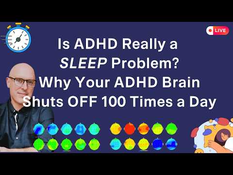
Is ADHD a Sleep Problem? Why Your ADHD Brain Shuts Off 100 Times a Day
Neurofeedback Works — But Here’s the Catch
Why You Feel Like Everyone Hates You (Even When They Don’t)
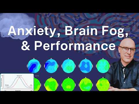
Stress: Anxiety, Brain Fog, & Performance
The Real Reason You're Anxious All the Time
Why Neurodivergent People Think Everyone Hates Them (Neuroscience Secret)
Why You Feel Broken… You Might Be Gifted and Tortured