ADHD Brain Types: Three Confirmed Patterns, One Diagnosis
If you carry an ADHD diagnosis, you got one of three labels: inattentive, hyperactive-impulsive, or combined. Those come from the DSM, a manual of behavioral checklists. They sort you into a bucket. They don't describe your brain.
Across 25,000 brain maps, the labels and the brains keep failing to line up. Two people with the same diagnosis can have completely different electrical patterns, respond to opposite interventions, and need different treatment plans. A 2025 structural imaging study put hard data under what brain-mapping practitioners have seen for years: ADHD is several distinct conditions wearing one name.
This guide pulls together what the imaging confirmed, what shows up in the QEEG, why sleep and anxiety travel with ADHD so reliably, and what stimulants and neurofeedback actually do to each pattern.
What did the 2025 study actually find?
Pan, Long, Quan and colleagues (JAMA Psychiatry, February 2025) used morphometric similarity network analysis. Instead of measuring activity at one location, this maps how structurally related regions co-organize across networks. Think of it as the structural cousin of QEEG connectivity work, where the useful signal lives in how regions relate, not just in how loud any single spot is.
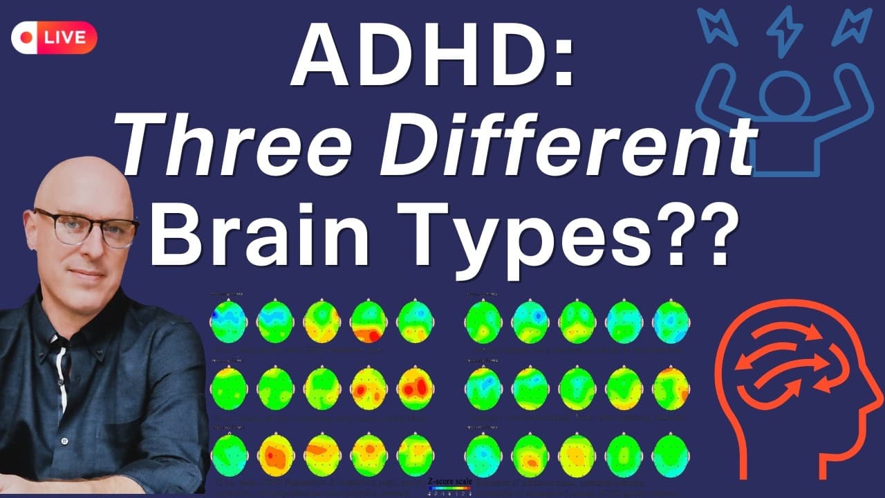
They found three biotypes, and they map cleanly onto patterns visible in the EEG:
Frontal slow activity (the inattentive brain). General frontal slowing, excess theta and alpha, high theta-to-beta and theta-to-alpha ratios. This is underactivation. The engine that won't start, won't stay in gear, won't pull through. The 1987 label "ADD" was actually a decent fit for this one.
Hyperarousal with excess fast activity (the hyperactive-impulsive brain). More right-sided, with heavy anterior cingulate involvement yoking into the pallidum. On the EEG you see excess beta, sometimes paradoxically fast and slow activity together. This is the disinhibited, fidgety, can't-pump-the-brakes presentation.
Emotional dysregulation (the third, underrecognized brain). A distinct structural pattern involving medial prefrontal cortex and pallidal networks, separate from the other two. The core problem is regulating emotional intensity, not sustaining attention or sitting still.
Shaw and colleagues (2014) argued emotional dysregulation should be treated as a core ADHD feature, mapping it to dorsolateral PFC and orbitofrontal cortex interacting with the amygdala and ventral striatum. Reward, threat, and danger-tagging circuitry. The 2025 biotype data backs that argument.
What does the third brain type feel like?
Rejection lands like a physical punch. Frustration escalates fast in school or at work. A small disappointment triggers a response that looks disproportionate from the outside. You may have been told you're "too much" or "too sensitive."
For decades this got filed as comorbidity: ADHD plus anxiety, ADHD with a mood problem. The biotype data says something different. For some people, the emotional reactivity is the ADHD, generated by the same dysregulated circuitry, not a separate condition stacked on top. This is where the "rejection sensitivity dysphoria" people describe online actually lives in the brain.
How do these three show up in a brain map?
The frontal-slow brain shows excess slow activity across the frontal and central strip, often with weak SMR (sensorimotor rhythm) ear to ear and low low-beta tone. One distinction most practitioners miss: frontal theta is not one thing. Midline frontal slowing is an anterior cingulate phenomenon, tied to error monitoring and selecting among competing thoughts. Pre-central slowing is about controlling and supervising behavior. Same "slow frontal lobe" label, two different generators, two different training targets.
The hyperactive-impulsive brain shows broken inhibitory tone, especially over the right inferior frontal gyrus and the right pre-central area. That right inferior frontal circuit signals down through the basal ganglia to pump the brakes. Without good control there, you can't stop the thing, can't stop saying the thing, can't stop fidgeting.
The emotional-dysregulation brain shows the cingulates and the pre/post-central gyri both sides involved. Picture a plus sign of tissue. Where the biggest pinches sit tells you where the work is. Front-midline beta runs with intrusive, obsessive thoughts. Front-midline theta runs with a looser, stimulus-seeking version: songs stuck in your head, a small tic, nail-biting.
These patterns are signposts, not verdicts. A feature that usually means anxiety makes it plausible you're anxious, not certain. People are weird. The map shows what's unusual for you compared to average. You and the clinician decide what's actually in your way. For the full picture of what a scan reveals, see QEEG Brain Mapping: What It Is, What It Shows, and What to Expect.
Further viewing: ADHD Isn't One Thing? Three Different Brain Types Confirmed.
Is ADHD actually a sleep problem?
Some researchers argue ADHD is fundamentally a sleep disorder, not an executive-function disorder. The overlap is real and mechanistic, not coincidental, even if that framing overstates the case.
Insomnia and delayed sleep-wake cycling are extremely common in ADHD. Biological markers like dim-light melatonin onset run later in this population. If your biological night ends late but school or work starts early, you get a chronic performance mismatch that looks exactly like inattention in the morning.
The mechanism runs through SMR, the beta-band rhythm over the central strip. When you're asleep, SMR has another name: sigma, or sleep spindles. Those spindles keep you asleep through a noise outside without forcing a full wake-up. The same C3 and central tissue that stabilizes attention while you're awake stabilizes sleep depth at night. Weak SMR means weak executive function and poor sleep maintenance, from the same circuit.
There's a second mechanism. Attention isn't continuously on; it browns out in micro-moments. Some of those brownouts look like fragments of sleep intruding into waking. Your ADHD brain effectively shuts off and reboots many times a day.
A 2025 study by Smullen and colleagues (Scientific Reports) found ADHD-like traits predict insomnia, and the mediator was pre-sleep cognitive arousal. The mind still scanning, rehearsing, refusing to hand off into sleep. The same regulation failure that won't keep you engaged in a task during the day won't let you disengage at night. The handoff doesn't complete in either direction.
The clinical implication: if you're training a brain for sleep and it carries significant ADHD features, go after the ADHD. For the broader sleep mechanics, see Biohacking Sleep: Optimize Your Rest for Peak Performance, and for the rhythm itself, SMR Neurofeedback: The Calm-Alert Brainwave That Trains Sleep, Focus, and Self-Control.
Further viewing: Is ADHD a Sleep Problem? Why Your ADHD Brain Shuts Off 100 Times a Day.
Why do ADHD and anxiety share a brain?
Anxiety disorders show up in roughly 25 to 50% of adults with ADHD. A 2026 paper by Bresser and colleagues (eClinicalMedicine) suggests the overlap runs deeper than comorbidity: "hyperarousal" is not one thing.
The team ran a factor analysis across questionnaires built for insomnia, anxiety, PTSD, ADHD, and depression, drawn from 467 adults. Hyperarousal broke into about seven dimensions: anxious arousal, somatic arousal, sensory sensitivity, sleep-related arousal, irritable arousal, vigilant arousal, and pseudomotor arousal (the flop-sweat, pounding-heart sympathetic surge).
These dimensions don't load equally across diagnoses. Insomnia carries a different hyperarousal profile than ADHD, which carries a different one than PTSD. Two people both saying "I'm stressed" can run completely different engines. One scans the environment for threat. The other can't shut off cognitive overactivation at bedtime. Same word, different shape, different intervention priority.
This connects back to the third biotype. The anterior cingulate that drives procrastination and stuck behavior also sits at the center of the emotionally reactive, low-inhibition presentation. ADHD and anxiety don't just co-occur. They share a driver.
On the EEG, hypervigilance shows as beta over the occipital tissue (O1, O2) with the eyes closed, or as a lack of alpha there. Fast alpha that runs more than half a standard deviation above average can make the mind race. Depleted or excessive delta tells you whether the system can downshift at all or keeps the throttle pinned.
For the anxiety side specifically, see Biohacking Anxiety: Targeting the Circuits That Won't Shut Up and Neurofeedback for Anxiety: What the Research Shows.
Further viewing: 7 Types of Hyperarousal: ADHD Into Anxiety.
Why don't 20 to 30% of people respond to stimulants?
Stimulants boost dopamine and norepinephrine, sharpen attention, and increase wakefulness. They follow an inverted-U: a little improves reaction time and sustained attention, more helps a bit, then you turn the corner and the dose throws you off.
On the map, ADHD medications typically reduce excess theta and raise weak low-beta toward a non-ADHD profile. Adults with ADHD show reduced cerebral blood flow in prefrontal and orbitofrontal regions; stimulants partly normalize that perfusion and network activation. For the frontal-slow brain, this works well. You can get strong activation, and neurofeedback that raises left-frontal beta and trims alpha excess pushes the same direction.
The non-responders are concentrated in the other two patterns. A hot front midline or low delta from poor sleep and stress means stimulants often produce more anxiety, more tics, intrusive thoughts, and worse sleep. That worse sleep feeds the dysregulation, and the whole thing spirals. The treatment didn't fail at random. It didn't match the biotype.
The alpha response is paradoxical and clinically useful. In an under-aroused, burnt-out brain, stimulants and caffeine raise alpha (more relaxed). In a well-rested brain, they lower it. I've mapped people with mixed anxiety-and-ADHD patterns who got an anxiolytic effect from a stimulant, with beta dropping rather than rising. The drug's effect depends on the brain it lands in.
One practical caution if you combine stimulants with neurofeedback: SMR and related protocols wash out tolerance. Three to four weeks in, a stable dose can start to feel like overmedication, with appetite suppression, sleep-onset trouble, and irritability. That's usually a good thing, an opening to step the dose down with a prescriber, but it surprises people who don't expect it.
Caffeine sits in its own category. It blocks adenosine rather than driving dopamine directly, carries genuine neuroprotective data across all-cause mortality, Parkinson's, and Alzheimer's, and rarely generates over-sensitivity. With a four-and-a-half-hour half-life clearing over roughly five half-lives, an afternoon cup can still be working at bedtime. For how stimulants intersect with dopamine and appetite circuits more broadly, see GLP-1 Drugs and the Brain: Ozempic, Dopamine, ADHD, Dementia.
Further viewing: What Stimulants REALLY Do to Your Brain (with QEEG Examples).
How do you match treatment to your brain type?
The three biotypes need three different approaches.
Frontal-slow / inattentive. Responds well to stimulants. Neurofeedback raises beta over the left frontal and central strip and reduces alpha excess. Sustained-focus meditation that anchors attention to a single object fits this brain. Address the sleep maintenance issue directly, because it's part of the same circuit.
Hyperactive-impulsive. SMR is the main lever. Build inhibitory tone over the central strip and right inferior frontal area. Meditation works better as a moving or active practice, because sitting on a cushion anchoring to nothing is the hardest possible task for this brain.
Emotional dysregulation. This is the pattern most often mismedicated. People here get treated for attention they don't primarily lack, or for anxiety and mood that are actually the ADHD. When the dysregulation runs severe with strong executive and mood features, some get labeled bipolar II. Map the cingulates and the pre/post-central gyri to find the biggest pinches. Anterior cingulate work helps the stuck, obsessive component. NAC tends to settle that tissue. Methylation analysis can matter here too: a quirky MTHFR or COMT genotype affects how these circuits run, and a targeted B-vitamin strategy can ease the cramp.
Neurofeedback takes time across every type. Resting EEG patterns shift slowly, roughly one standard-deviation color shade and about 15 points on an attention test by 20 to 30 sessions, with stable change in anxiety and ADHD features closer to 40 to 50. Track progress by day-to-day subjective change confirmed later by the map and an attention test (the IVA-2 is what I use), not by within-session trend lines, which are too noisy to trust.
For where the evidence stands, see Does Neurofeedback Work for ADHD? A Neuroscientist's Guide and Is Neurofeedback Legitimate? A Research Overview.
A note on evidence strength
The 2025 biotype study confirms what EEG phenotype research has shown for years (Clarke's 2001 subtypes, Monastra's theta-beta work, Slater's 2022 review), but it does it in JAMA Psychiatry with large-scale imaging. That changes who pays attention: psychiatrists and parents, not just the brain-mapping back rooms.
The hyperarousal-dimensions and ADHD-as-sleep findings are newer and less settled. A 2025 preprint by McCormick points in a different direction with different measures. Neurotransmitter claims stay inferential; we don't have clean human studies on "chemical imbalance," and that language is mostly marketing.
What holds up: ADHD is several distinct conditions. The label sorts behavior; the brain runs the show. Map your brain before you choose an intervention, find which of the three patterns is your bottleneck, and match the tool to the circuit. The next concrete step is a QEEG, which costs less than a month of guessing wrong with the wrong medication.
TAGS
References
- Shaw (2014). Emotion Dysregulation in Attention Deficit Hyperactivity Disorder. doi:10.1176/appi.ajp.2013.13070966
- Smullen (2025). Pre-sleep arousal as a possible mechanism driving sleep problems in relation to ADHD traits. doi:10.1038/s41598-025-09866-3
Get new articles and brain training insights by email.
No spam, unsubscribe anytime.
Related Articles
Why Won't Your Brain Shut Up? The Neuroscience
Racing thoughts, 3am anxiety, and a mind that won't quiet down are circuits, not character flaws. A clinical neuroscientist maps the phenotypes behind it.
QEEG Brain Mapping: What It Is, What It Shows, and What to Expect
A neuroscientist explains what QEEG brain mapping measures, how it differs from a hospital EEG, what the maps show, and what happens during an assessment.
Biohacking with EEG Phenotypes: Predicting Brain Function from Electrical Patterns
EEG phenotypes are stable electrical signatures that predict how you process attention, regulate mood, and respond to neurofeedback and medication.
About Dr. Andrew Hill
Dr. Andrew Hill is a neuroscientist and pioneer in the field of brain optimization. With decades of experience in neurofeedback and cognitive enhancement, he bridges cutting-edge research with practical applications for peak performance.
Get Brain Coaching from Dr. Hill →