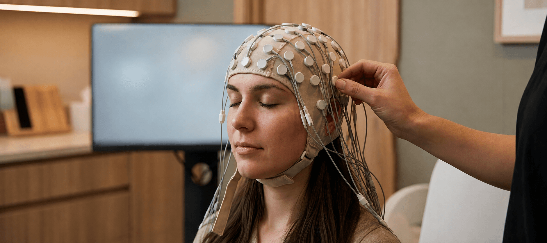
A QEEG brain map is a quantitative analysis of an EEG recording. It can summarize features of scalp electrical activity and compare selected measures with a reference dataset. The colors make differences easy to see. Understanding what produced a difference takes more work.
Begin with the recording. EEG measures electrical activity through electrodes placed on the scalp. Movement, alertness and medicines can affect the result. The MedlinePlus EEG guide explains these considerations and advises following preparation instructions without changing medication independently.
The map is downstream of the raw signal
A quantitative result depends on the data selected for analysis. A segment affected by movement or muscle activity can distort a measure if it is treated as brain activity. Eyes-open and eyes-closed recordings also answer different questions about the state being sampled.
Ask how the raw EEG is inspected, how unusable segments are handled and which conditions were recorded. Ask what reference data and analysis methods the report uses. A detailed image is only as informative as the recording and the choices behind it.
Comparing a value with an age-matched distribution can tell you how unusual that value is under the comparison used. It leaves a further question about whether the difference explains a difficulty, matters to functioning or predicts a useful intervention.
Consider a theta/beta power ratio, a measure discussed in the American Academy of Neurology’s advisory. The calculation divides power in a slower frequency band, theta, by power in a faster band, beta. It turns two measurements from the recording into a single comparison. A higher ratio can result from more theta power, less beta power, or both, so the ingredients matter as well as the ratio.
Now imagine an illustrative report whose legend assigns red to ratios above its reference range. Red would encode that comparison under that report’s rules. To understand it, you would still need the recording condition, the underlying measurements and the reference being used. The color alone supplies no measure of how difficult homework or sleep feels. This is an example of reading a display, with no patient data or universal color scale implied.
Different uses require different evidence
The FDA-NIH biomarker framework distinguishes uses such as diagnosis, prediction and monitoring. A measure useful for observing change is not automatically validated for selecting a treatment or diagnosing a condition.
For example, the American Academy of Neurology's theta/beta advisory cautions against using that ratio to confirm ADHD outside research or replace a clinical evaluation. This is a specific boundary for a specific proposed use. It should neither be ignored nor expanded into a claim about every EEG application.
The clinical question comes first. What does the person need help understanding, and would this measurement change a decision in a defensible way? If the same training would be prescribed regardless of the result, ask what the map is contributing.
Put the result beside the history
Sleep, symptoms, functioning and the circumstances of the recording belong in the interpretation. A person can recognize a vivid description attached to a map while the causal claim remains uncertain. Familiarity with the description is a starting point for discussion, rather than validation of the biological explanation.
An attention test can add another kind of information. Agreement between two measurements may be interesting, but it still needs an appropriate interpretation. Neither should be made to answer a question it was never validated to answer.
The EEG phenotype article examines how recurring patterns become research hypotheses. The ADHD brain-types article distinguishes those patterns from clusters found in MRI research.
Questions to bring to a consultation
Ask what the report can establish, what remains uncertain and how alternative explanations are considered. Request an explanation in ordinary language of any finding that would change a recommendation.
If training follows, agree on an outcome outside the map and a review process. A change in the map can be recorded alongside changes in daily function; neither should be silently substituted for the other. Appropriate medical concerns should be directed to a qualified clinician.
QEEG can make electrical patterns visible and comparable. Its value depends on keeping the chain of reasoning open to inspection: the signal, the analysis, the interpretation and the decision. Every link deserves a clear explanation.
References
Get new articles and brain training insights by email.
No spam, unsubscribe anytime.
Related Articles
Is Neurofeedback Legitimate? Evidence and Limitations
Examine how neurofeedback works, what controlled studies show, and the questions that help distinguish responsible practice from unsupported promises.
Remote Neurofeedback: How Home Training Works
Understand home neurofeedback equipment, supervision, recording quality and practical commitments before deciding whether remote training suits you.
Neurofeedback Cost: Prices, Packages and Questions
Compare neurofeedback program costs, understand what a package includes, and ask useful questions about reassessment, refunds and possible non-response.
About Dr. Andrew Hill
Dr. Andrew Hill is a neuroscientist, founder of Peak Brain Institute and host of the Head First podcast. He writes about neurofeedback, attention, learning and brain health.
Get Brain Coaching from Dr. Hill →